

Neurons are highly organized cells that are specialized to communicate information.
Estimate: 100-1000 billion neurons in human nervous system.
Each part of the neuron plays a specific role in this communication.
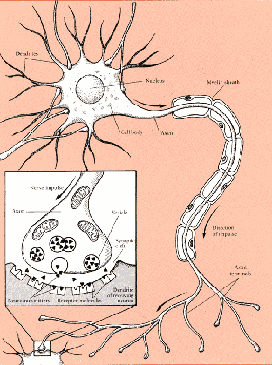
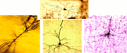
Variable shape and size Contains nucleus. Functions: metabolic machinery of cell (i.e., Protein Synthesis, Metabolism,Energy Production, etc.)
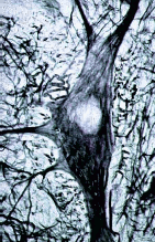
site where the axon exits the soma. Functions: establishing the action potential
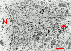
single long thin extension, (branched in some neurons), constant diameter
Function: electrochemical transmission - sometimes covered with myelin.
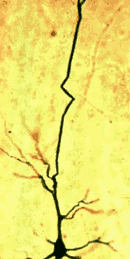

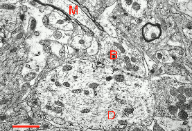
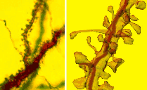
thin branching fibers, spiny or aspiny, decreasing diameter. Functions: inputs- reception of transmissions from adjacent neurons.
Electrochemical process
Propagation of impulse within neuron (electrical).
Transmission of impulse between neurons (chemical).
Resting Potential
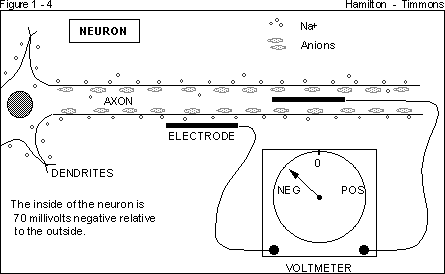
Neuron at rest.
Not firing.
Stable, negative charge inside neuron relative to outside (- 70 mV)
(Hyperpolarization)
Where does the resting potential come from?
1. Elemental ions:
K+ (potasium)
Na+ (sodium)
CA++ (calcium)
Cl- (chloride)
2. Elemental forces:
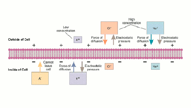
diffusion / concentration gradient
electrical potential (voltage) / electrostatic gradient
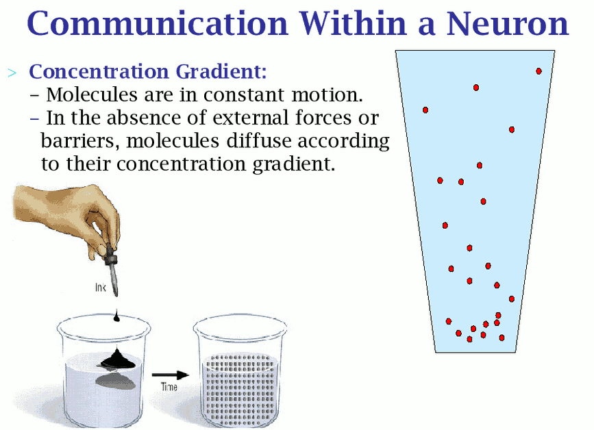
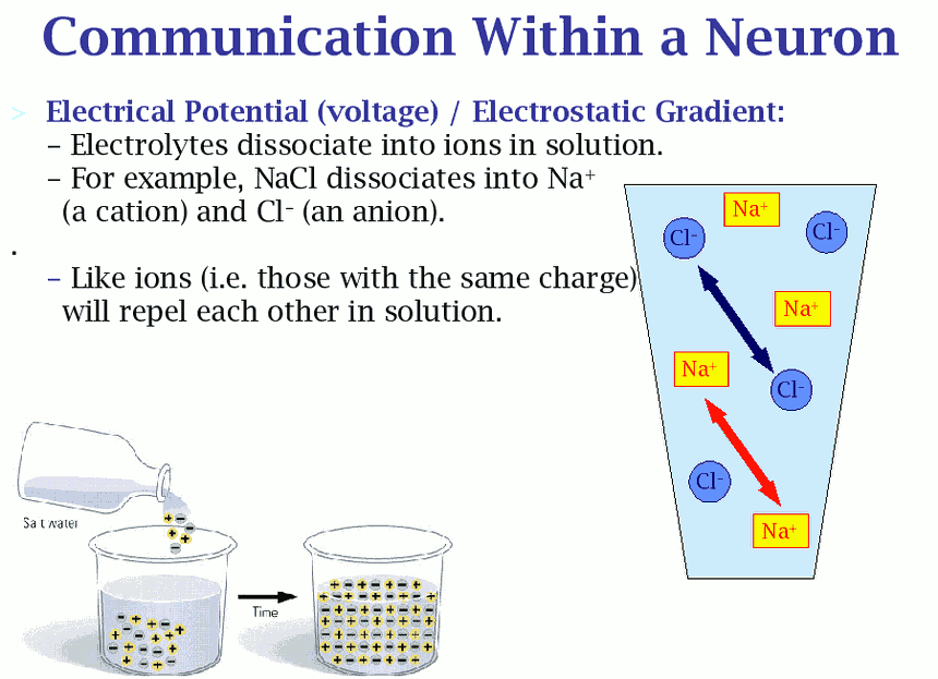
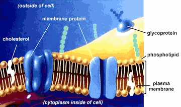
cell membrane at rest is largely impermeable to Na+ ions
although ion channels or ionophores allow some ions
through
Na/K ion pump – moves 3 Na+ outside moves 2 K+ inside
results in: more (+) ions outside than inside.
the membrane permeability determines resting potential through 2 types of ion channels:
1. gated – let certain ions through under certain circumstances.
2. Non gated – always lets certain ions through, but not fast enough to neutralize the ion pumps.
Action Potential
Neuron receives sufficient stimulation from receptor, other neurons, etc.
Na+ ions rush in.
Interior of axon becomes positive relative to the outside (+ 40 mV)
(Depolarization)
This release of energy passes down the axon and is the firing of the neuron.
Once neuron fires, resting potential returns.
All or None Law
Once neuron fires, the impulse moves down the axon at the same intensity.
No stopping it.
Impulse frequency
While there are differences in speed of transmission between neurons, increase in neuron stimulation doesn’t cause stronger impulse. Speed and electrical change remains the same regardless of intensity.
Stronger stimulation leads to more frequent firing of the neuron or more neurons activated.
Absolute Refractory Period
Brief period of time after firing when neuron won’t fire no matter how much stimulation given.
As brief as 1/1000 of a second. HOWEVER, means a limit as to how many times neuron can fire per
second (the maximum is 500-600 times/second).
Think of neuron like a toilet that works well
Information flows in 1 direction (out to the sewage!)
It is all or none - the toilet doesn't have "half flush" days
There is a period of time that, no matter how much you jiggle the handle, no flushing will occur
Depolarization – resting potential decreases (towards zero and positive numbers) hyperpolarization - resting potential increases (larger negative numbers)
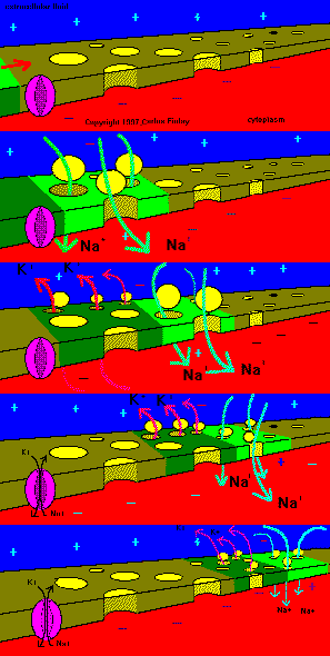
When depolarized past the threshold, the resting potential breaks down.
Voltage-gated Na+ ion channels open and ions flow in along diffusion and electrostatic gradients.
K+ channels then open allowing K ions to flow out.
Gates close and ion pumps restore normal resting potential.
All-or-None Law:
All action potentials are the same. You either have
one or you don’t.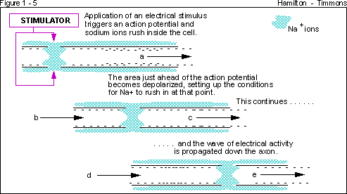
Post synaptic potentials are generated on dendrites ans cell bodies
Action potentials move along the membranes of a neuron. They are initiated at the axon hillock and travel along axons away from the cell body.
Where do action potentials come from naturally?
Sensory neurons
They are generated in cell bodies which depolarize in response to specific stimulation. Called Post Synaptic Potentials (PSP).
The more depolarization, the faster action potentials are generated at the axon hillock.
Neurons do not touch each other
How does activity in one neuron influence activity in another?
Synapses
Presynaptic neuron:
• terminal bouton at the end of axons
• synaptic cleft
Post synaptic neuron
-receptor sites
Activity at synapses
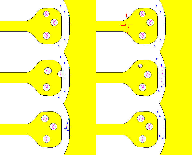
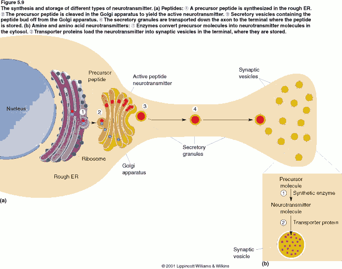
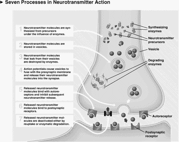
The arrival of an action potential at terminal causes C++ ion channels to open. This causes the release of a neurotransmitter from synaptic vesicles into the synaptic cleft.
The neurotransmitter interacts with receptor sites located on the surface of large protein molecules. This causes a change in the resting potential of the membrane of the cell body of the post synaptic cell by opening ion channels .
May directly (gated ion channel) or indirectly (second messenger) open ion channels.
Depolarization causes excitation (opens Na+ channels) and increases firing rate. (Excitatory post synaptic potential, EPSP)
Hyperpolarization causes inhibition (opens Cl-
channels) and decreases firing rate. (Inhibitory post synaptic
potential, IPSP)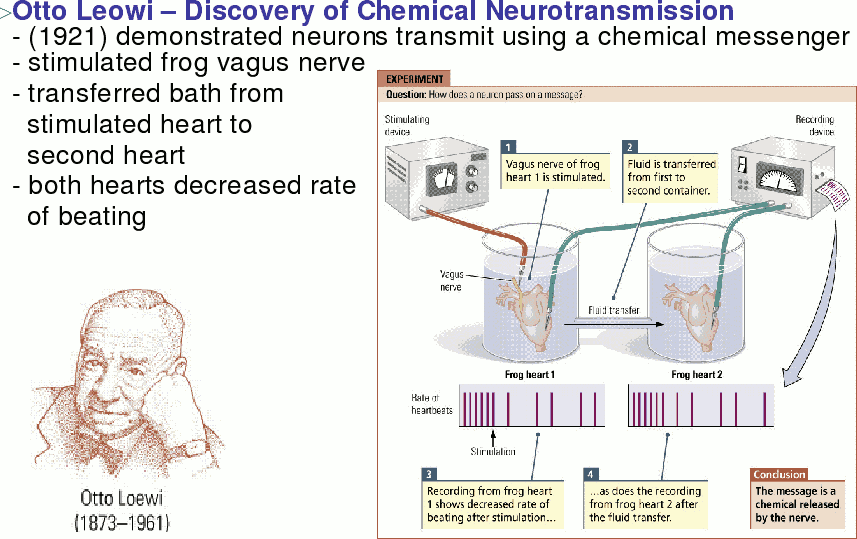
Summation of excitation and inhibition
• spatial
• temporal
Slow, long acting changes
Second messengers
• may open ion channels
• may alter other aspects of cell functioning
Long term changes
Kinases – Longer acting than second messengers
Permanent changes
Transcription factors – alter the expression of DNA and can change the production of ion pumps, ion channels and even synapses.
Transmitter action terminated by reuptake or enzymes.
Neuromodulator: cause changes in reactivity of postsynaptic membrane to other neurotransmitters.
May act internally, or externally
Neurotransmitters
Acetylcholine
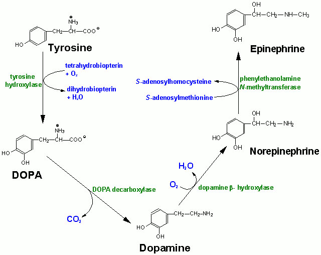
Monoamines (biogenic amines)
--catecholamines
--Norepinephrine (NE)
--Dopamine (DA)
--Epinephrine (E)
--Indoleamine
--Serotonin (5-HT)
Amino acids:
GABA
Glycine
Glutamate
Peptides:
--Substance P
--endogenous opiates (mrphinelike substances)
--enkephalins
--endorphins
various hormones
• somatostatin
• vasopressin
Dale’s Law: neurons always produce the same neurotransmitter (s) at all synapses.
Cholinergic, dopaminergic, serotonergic, etc
The effect of a transmitter depends on the nature of the receptor. Each transmitter may have many types of receptor.
Level of neurotransmitter release can be governed by autoreceptors on the presynaptic membrane
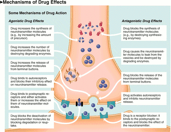
Drugs interfere with neurotransmission by interfering
with what happens at synapses can block or facilitate.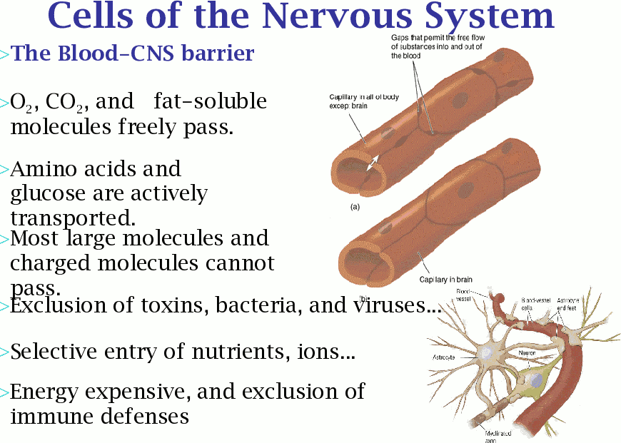
Acetylcholine (Ach):
deactivated by acetylcholinesterase (AchE)
Acetylcholinesterase inhibitors:
• organic phosphates – insecticides like Maticil
• nerve gasses like sarin
two types of Ach receptor sites:
stimulated blocked
muscarinic muscarine atropine
nicotinic nicotine curare
Monoamines:
• Norepinephrine
• Epinephrine
• Dopamine
• Serotonin
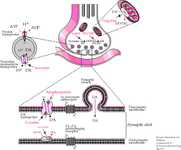
Destroyed by two enzymes:
Monoamine Oxidase (MAO)
COMT
Reuptake – blocked by cocaine – SSRIs
Receptor subtypes:
E and NE:
alpha-1 alpha-2 beta-1 beta-2
Dopamine:
D1........D6
Serotonin:
5-HT1 (4 subtypes).....5-HT4
Gamma-aminobutyric Acid (GABA)
universally inhibitory – linked to a Cl- ion channel.
Glutamate:
excitatory transmitter
NMDA receptor
non NMDA receptor
Peptides:
endorphins – 16-30 amino acids
enkephalins – 5 amino acids
receptor types:
mu, kappa and delta receptors
blocked by nalorphine
The nervous system
CNS – brain and spinal cord
PNS – everything else
bundles of axons – white matter nerves (PNS) or tracts (CNS) clusters of cell bodies - grey matter
ganglia (PNS) or centres (nuclei) (PNS) Peripheral Nervous system (PNS) somatic:
-----conscious senses
-----voluntary motor
autonomic:
-----sympathetic NS – fight-or-flight – epinephrine
-----parasympathetic NS – vegetative functions - ACh Central Nervous system (CNS) spinal cord
relay center
reflexes
ventral – motor output
dorsal – sensory input
Brain
100,000,000,000 neurons each with synapses on 1000 other neurons and 10,000 synapses from other neurons
medulla – autonomic control
• breathing
• vomiting
RAS and Raphé System
RAS – arousal
Raphé System
• sleep
• mood
Locus Coeruleus
mood: fear, panic, anger
primarily NE,
stimulated by monoamines
inhibited by GABA
Cerebellum:
coordination of motor control
Basal Ganglia:
striatum – input from thalamus and cortex
globus palladus – output side with feedback to thalamus
coordination of motor control
• - -extrapyramidal motor system
• - -DA receptors
• - -DA deficiency – Parkinsons Disease
Periaquiductal gray:
pain control – mu receptors and morphinelike transmitters punishment system
Limbic system:
hypothalamus – eating and drinking control
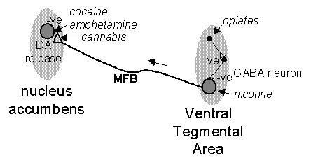
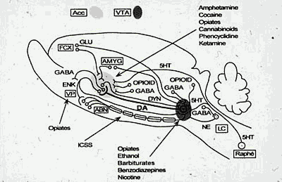
medial forebrain bundle
--- reinforcement centres
---mesolimbic system (DA)
------ ventral tegmental area
------nucleus accumbens
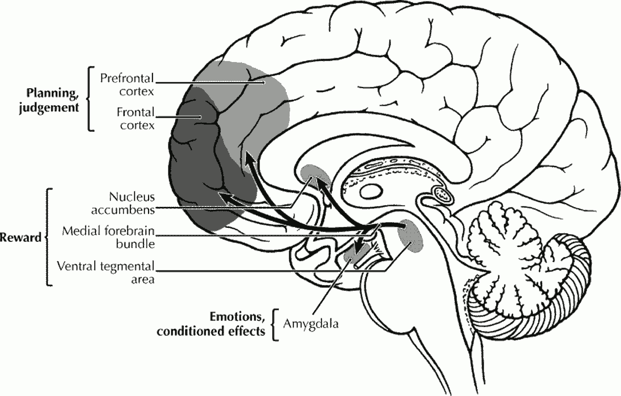
hippocampus – leraning and memory
amygdala and septum – serotonergic input from the Raphé system Aggression and emotion
Cortex
sensory input areas
motor control output areas
language
memory and thinking
glutamate – excitatory transmitter
GABA – inhibitory transmitter
Development of the Nervous system
cells formed during first 12 weeks
migrate to appropriate location
send out axons to correct target
form synapses
controlled by chemical signals
psychoactive drugs can disrupt the process because:
1. get into the brain
2. confuse or block chemical signals
Teratology – brain malformation, eg,
Fetal Alcohol Syndrome
Functional or behavioral teratology: there may be no anatomical malformations, but functioning of the brain may be disturbed. There may be behavioral changes.